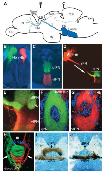
Figure 1. Laterotopic Projections from the Habenular Nuclei to the IPN.
(A) Schematic illustration of a lateral view of an adult zebrafish brain showing the telencephalo-habenulo-interpeduncular pathway that connects the telencephalon with the ventral midbrain.
(B–G) Confocal images of sections of brains of adult fish in which left habenular axons are anterogradely labeled with DiO (green) and right habenular axons are labeled with DiI (red).
(B and C) Transverse sections at the level of the habenulae (B) and the IPN (C) showing left habenular axons (green) terminating in the dorsal IPN and right habenular axons (red) terminating in the ventral IPN.
(D and E) Low- (D) and high (E)-magnification views of a parasagittal section showing axonal projections from the right habenula (red) through the FR to the IPN. Within the IPN, dorsal and intermediate regions (green) are predominantly innervated by axons from the left habenula.
(F and G) Horizontal sections showing projections from the left habenula (green) terminating in the intermediate IPN (F) and projections from the right habenula (red) terminating in the ventral IPN (G). In both cases, the labeled axons surround the fluorescent Nissl-stained cell bodies (blue) at the center of the nucleus.
(H) Dorsal view of the intact ventral midbrain in a 5-day-postfertilization (dpf) larva, showing a 3D reconstruction of projections from the left (green) and right (red) habenulae. The oculomotor nucleus is visualized with a foxD3:GFP transgene (blue). Some FR axons on both left and right sides bypass the IPN and terminate more caudally (arrows).
(I and J) Transverse sections through the IPN of 4-dpf larvae showing projections from the left (I) and right (J) habenulae labeled with photoconverted DiI.

