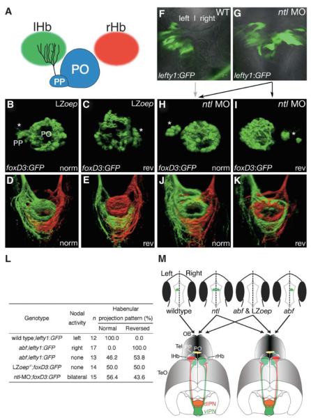
Figure 4. Laterotopic Habenular Projections Are Still Established in Embryos Laking Lateralized Nodal Activity in the Epithalamus.
(A) A cartoon viewing dorsally onto the epithalamus showing the parapineal organ innervating the left habenula. (B–K) LZoep−/−;Tg(foxD3:GFP) (B–E), wild-type (F), and ntl morphant (G–K) embryos and larvae in which Nodal-pathway activity (F and G) or parapineal (asterisks) laterality (B, C, H, I) was assessed by GFP-transgene (indicated bottom left) expression and left and right habenular projections were subsequently assessed by DiI and DiA labeling at 4 dpf (D, E, J, and K). LZoep−/− embryos show an equal number of cases of parapineal migration to the left (B) and to the right (C). Position of the parapineal is concordant with the orientation of laterotopic innervation of the IPN (D and E). The left-sided activation of lefty1:GFP in the wild-type embryo (F) is normally associated with subsequent migration of the parapineal organ to the same side of the brain (indicated by the gray arrow). lefty1:GFP is activated bilaterally in ntl morphants (G), leading to randomization of parapineal position (black arrows to [H] and [I]), which is 100% concordant with the orientation of laterotopic habenular connectivity (J and K). (L) Orientation of laterotopic habenular projections in mutants and morphants that are defective in the expression of Nodal-pathway genes in the epithalamus. Habenular projection patterns were assessed in 4-dpf larvae by carbocyanine-dye axon tracing. The orientation of laterotopy is described as “normal” when left habenular neurons innervate the dorsal IPN and as “reversed” when right-sided axons terminate in the dorsal IPN. (M) Nodal activity determines the orientation of laterotopy in habenulo-interpeduncular projections. The top panels are cartoons of dorsal views of late-somite-stage embryos showing activation of Nodal signaling (green) in the epithalamus. The bottom panels show sub-nuclear organization of the habenulae and laterotopic projections from the habenulae to the IPN in normal and reversed-laterality adult fish. In wild-type embryos, we propose that Nodal activity in the left epithalamus indirectly leads to expansion of the lateral sub-nucleus (red) in the left habenula and the medial sub-nucleus (green) in the right habenula, resulting in projection of the left habenula predominantly to the dorsal IPN and of the right habenula to the ventral IPN. This pattern of sub-nuclear organization is completely reversed (right lower panel) in those abf mutant embryos in which Nodal signaling is activated on the right side. When Nodal signaling is activated bilaterally (ntl) or is absent (abf; LZoep), habenular asymmetry is still established, but laterality of the sub-nuclear organization and consequently of the laterotopic innervation of the IPN becomes randomized.
Abbreviations: norm, normal laterality; PO, pineal organ; PP, parapineal; and rev, reversed laterality.

