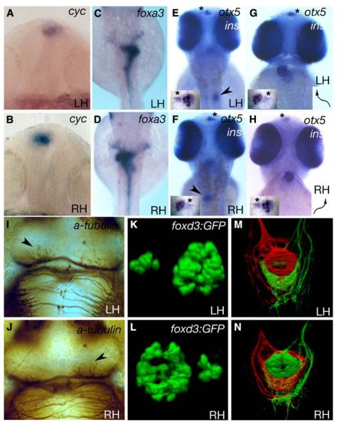
Figure 1.
Some fsi Embryos Show Complete Situs Inversus
Frontal (A, B, G, and H) and dorsal (C–F, I–N, and insets in [E]–[H]) views of LH and RH fsi fry.
(A and B) Epithalamic expression of cyc.
(C and D) Expression of foxa3 in the gut.
(E and F) insulin (pancreas, arrowheads in E,F) and otx5 expression (pineal and parapineal; inset, otx5 expression; asterisks indicate parapineal position).
(G and H) cmlc2a expression in the heart and otx5 in the pineal and parapineal nuclei (arrows indicate direction of heart looping; inset, otx5 expression; asterisks indicate the position of the parapineal nucleus). Note that the reversed position of the parapineal nucleus in E and G (LH) compared to F and H (RH) is due to different perspectives (dorsal versus frontal views). (I) and (J) show anti-acetylated tubulin labeling of the habenular neuropil (arrowheads indicate the nucleus with more robust labeling).
(K and L) 3D reconstructions of pineal (large nucleus) and parapineal (small nucleus) expression of a foxd3:GFP transgene.
In all cases, LH and RH fsi fry show opposite laterality of structures. ([A and B] 18–20 somites; [C–J] 2.5 dpf).
(M and N) Dorsal view of the intact ventral midbrain in 4 dpf LH and RH fsi fry; shown is a 3D reconstruction of projections from the left (red) and right (green) habenulae within the IPN. There is a DV inversion of projection patterns in the RH fsi fry.

