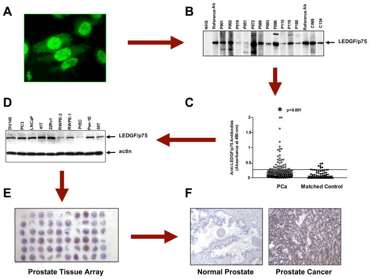
Fig. 1. The path to the identification of LEDGF/p75 as a candidate TAA in prostate cancer.
A, autoantibodies with a nuclear fluorescence staining pattern resembling that of the known autoantigen LEDGF/p75 (also called DFS70) were initially detected in prostate cancer sera by screening the sera in Hep-2 slides and LnCaP prostate cancer cells growing on coverslips via immunofluorescence microscopy. B, the presence of these autoantibodies in prostate cancer sera (p) and some positive matched control sera (c) was detected by immunoblotting of LnCaP cell lysates using reference anti-LEDGF/p75 sera as positive controls. C, the frequency of these autoantibodies in prostate cancer was determined in an ELISA system using purified recombinant LEDGF/p75 by testing against sera from prostate cancer patients and age- and gender-matched controls. Significantly high frequencies were obtained in the prostate cancer group. D, validation of LEDGF/p75 as a protein associated with prostate cancer was first achieved by analyzing its expression in a panel of prostate cancer cell lines by immunoblotting. High expression was observed in most tumor and transformed cell lines. The lowest expression was in the primary normal cell line PrEC. E, the availability of the human prostate cancer Histo-Array™ facilitated immunohistochemical analysis of LEDGF/p75 in tumor tissue. F, LEDGF/p75 was found to be highly expressed in the majority of prostate tumor tissue specimens but not in normal prostate tissue. For details see Ref. 27.

