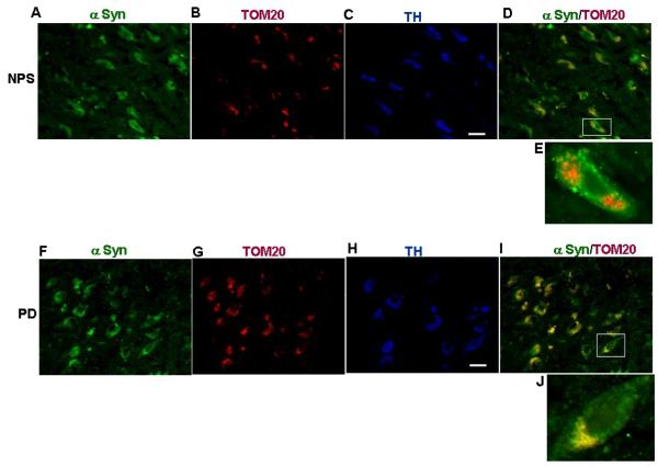
Figure 2. Immunofluorescence microscopy analysis of mitochondrial α synuclein in dopaminergic neurons of substantia nigra of non-PD and PD subjects.
Figures A-E: Triple labeling of dopaminergic neurons of substantia nigra of post mortem non-PD subject (NPS # 11) with anti rabbit α synuclein (A), anti goatTOM20 (B), and anti mouse TH (C). (D) Merged image of A and B. E= enlarged neuron. Figures F-J: Triple labeling of dopaminergic neurons of substantia nigra of post mortem PD subject (#11) with anti rabbit α synuclein (F), anti goatTOM20 (G), and anti mouse TH (H). (I) Merged image of F and G. J= enlarged neuron. Immunostaining was carried out as described in ref# 56. Bar= 100 μm.

