
Figure 2.
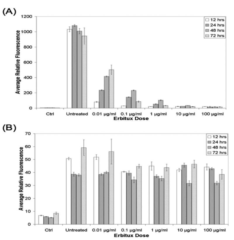
The EGF uptake of the (A) U251-GFP cells and (B) 9L-GFP cells as measured by EGF-AF647 fluorescence, following varied incubation time with difference concentrations of cetuximab. Ctrl indicated fluorescence for cells that were not incubated with EGF-AF647 to show the background signal at the excitation and emission wavelength used to measure the EGF-AF647 fluorescence. Untreated indicated the EGF-AF647 fluorescence signal from cells that were not treated with cetuximab therapy. Each bar represents the average of three samples and the error bars show the standard deviation of the mean.