
Figure 3.
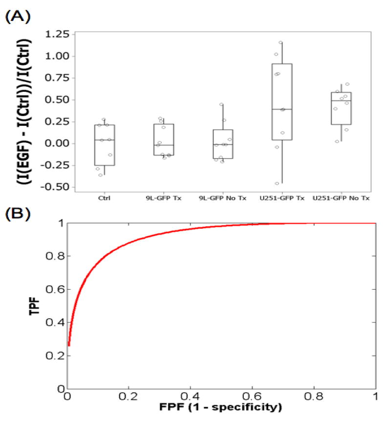
(A) The EGF-IRDye fluorescence on day 14 of the experiment for the treated and untreated U251-GFP and 9L-GFP groups as well as the control group. (B) Untreated EGFR+ U251-GFP and EGFR− 9L-GFP tumors could be stratified by ROC analysis with an AUC = 0.92.