
Figure 4.
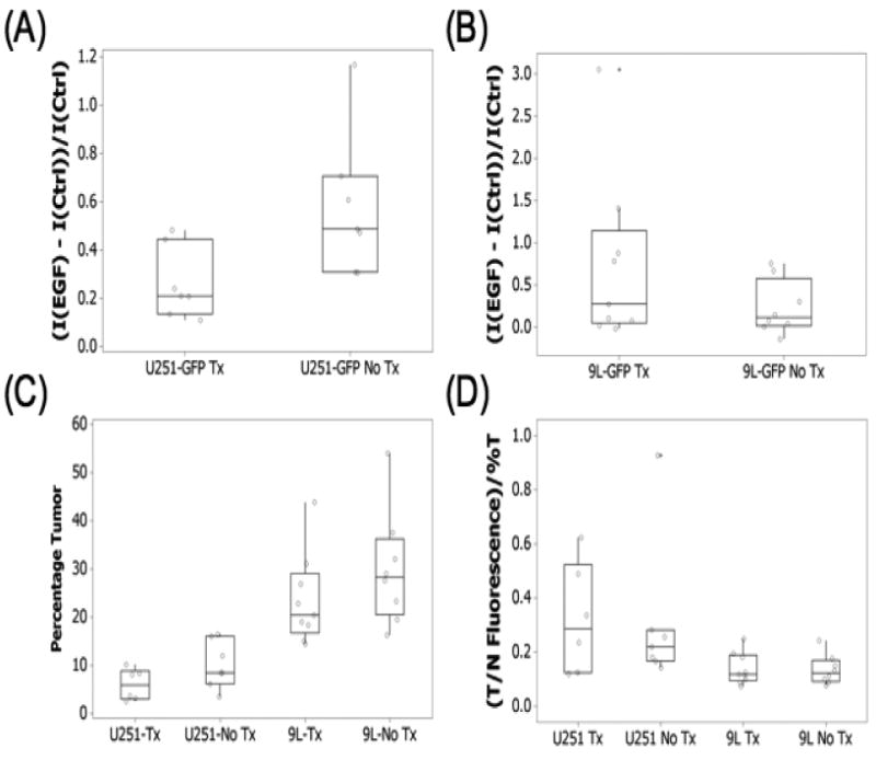
The EGF-IRDye fluorescence during the second week of the experiment, 24 hours after administration (day 22) for the treated and untreated (A) U251-GFP and (B) 9L-GFP tumor-bearing animals. The mice were sacrificed on day 24 of the experiment and their brains analyzed by (C) GFP fluorescence for tumor size, which is shown as a percentage of the brain and (D) EGF-IRDye fluorescence. The EGF-IRDye tumor to brain tissue fluorescence was normalized to the tumors size as determined by GFP fluorescence.