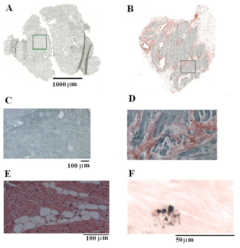
Figure 5.

LA fibrosis, fatty deposits and iron deposits in patients undergoing cardiac surgery. A and B, montage images of atrial tissue after staining with picrosirius red (fibrosis-red, muscle-green). A: LA biopsy from a 30-year old patient who remained in normal sinus rhythm shows no evidence of fibrosis. B: LA biopsy from a 74 year old patient who developed atrial fibrillation shows 16% atrial fibrosis. C: Enlarged (10X) boxed region in A, demonstrates a lack of fibrosis. D: Enlarged (10x) boxed region in B demonstrates the highest amount of fibrosis. E: 20X left atrial sample stained with hematoxylin and eosin demonstrating fatty infiltration (white circles) within the muscle section. F: 40X left atrial sample stained with modified Perls demonstrating atrial muscle (pink) and iron deposition (blue).
