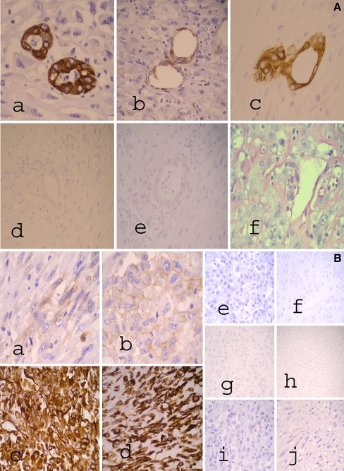
Fig. 3.
A Cholangiocarcinoma component with immunohistochemical and Mucicarmine stains showing (a) CK7, (b) CD56, and (c) CK19 positivity, (d) Hepar-1, and (e) Glypican-3 negativity, and (f) Mucicarmine stain showing cytoplasmic globules. B Sarcomatous component with selected immunohistochemical stains for epithelioid and spindle-cell elements. In (a), (b) CD56 shows focal weak positivity in both (a) spindle and (b) epithelioid cell elements. In (c), (d) Vimentin is strongly positive in both (c) epithelioid and (d) spindle cell elements. For epithelioid cell elements, (e) CK7 and (f) CK19 are negative, (g) Hepar-1 and (h) Glypican-3 are also negative, and (i) CD117 is negative. For spindle-cell elements, (j) CD117 is negative

