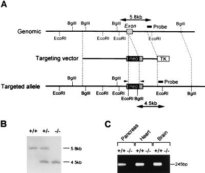
Figure 1.
(A) Schematic representation of the mouse Ki6.2 gene, targeting vector, and targeted allele. The exon is indicated by shaded boxes. Neo and TK indicate a neomycin-resistant gene and a herpes simplex virus thymidine kinase gene, respectively. Restriction sites are indicated. The probe used for Southern blot analysis is shown. Primers used for reverse transcription–PCR (RT-PCR) are indicated by arrowheads. (B) Southern blot analysis of F2 offspring. Genomic DNA was digested with EcoRI and BglII and hybridized with the probe. Lanes: +/+, wild type; +/−, heterozygote; −/−, homozygote. (C) RT-PCR analysis of pancreas, heart, and brain of Kir6.2+/+ and Kir6.2−/−. cDNAs were synthesized from total RNA (10 μg) from the tissues of Kir6.2+/+ and Kir6.2−/−. The expected size of the PCR product (245 bp) is indicated. Lanes: +/+, heterozygote; −/−, homozygote. The Kir6.2 transcript was not detected in tissues of Kir6.2−/−.

