Figure 2.
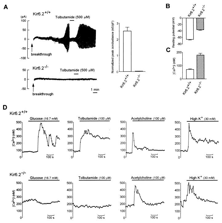
(A Left) Representative traces of whole-cell recordings of pancreatic beta cells in Kir6.2+/+ and Kir6.2−/−. The holding potential was −70 mV, and alternate voltage pulses of ±10 mV and 200-ms duration every 2 s were applied. In Kir6.2+/+ beta cells (Upper), dialysis of the beta cells intracellularly with the ATP-free pipette solution (breakthrough) caused a progressive increase in K+ conductance, and addition of 500 μM tolbutamide promptly inhibited this conductance. In contrast, no increase in K+ conductance was observed in Kir6.2−/− beta cells (Lower). (A Right) Normalized peak KATP channel conductance of pancreatic beta cells in Kir6.2+/+ and Kir6.2−/−. Since the membrane area of each beta cell varied, ATP-sensitive conductance was normalized by dividing by the membrane capacitance for each cell. The KATP channel conductance in Kir6.2+/+ beta cells was 2.50 ± 0.25 nS/pF (n = 36) and was completely lost in Kir6.2−/− (0 nS/pF, n = 38). (B) Resting membrane potential of Kir6.2+/+ and Kir6.2−/− beta cells. (C) Basal [Ca2+]i in Kir6.2+/+ and Kir6.2−/− beta cells. (D) Effects of various insulin secretagogues on [Ca2+]i in Kir6.2+/+ and Kir6.2−/− beta cells. The secretagogues used were glucose (16.7 mM), tolbutamide (100 μM), acetylcholine (100 μM), and K+ (30 mM). In Kir6.2+/+ beta cells, these secretagogues all increased [Ca2+]i. In contrast, in Kir6.2−/− beta cells, acetylcholine or K+ increased [Ca2+]i, but neither glucose nor tolbutamide increased [Ca2+]i. Horizontal bars indicate application periods of the agents. Representative examples are shown.

