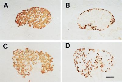
Figure 4.
Histology of pancreatic islets in Kir6.2+/+ and Kir6.2−/−. Pancreatic beta cells (A and C) and alpha cells (B and D) were stained by using guinea pig antiinsulin and rabbit antiglucagon antibodies, respectively, as described previously (25). In the islets of Kir6.2−/−, glucagon-positive alpha cells (D), which are present in the periphery of the islets of Kir6.2+/+ (B), are seen also in the central region of the islets. The beta cell population in Kir6.2−/− (C) is not different from that in Kir6.2+/+ (A). (Bar = 100 μm.)

