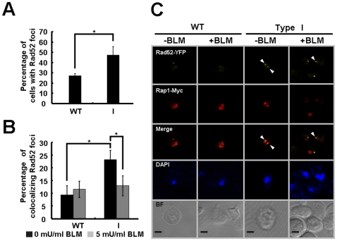
Figure 6. Localization of Rad52 to telomeres in cells with short telomeres.
(A) The percentage of cells forming spontaneous Rad52-YFP foci was scored as described in Materials and Methods. (B) The percentage of Rad52-YFP foci colocalized with Rap1-Myc. Wild-type and type I cells expressing Rad52-YFP and Myc-tagged Rap1 were arrested in G1 with α-factor for 2.5 hours. Half of the culture was exposed to bleomycin for 30 min to generate DSBs. Cell treated with or without bleomycin were released into S-phase for 90 min. Before the addition of belomycin, the proportion of colocalized Rad52 in type I cells differed from that in wild-type in a statistically significant manner (P values = 0.019), and in type I cells, the percentage of colocalization prior to bleomycin treatment (black bars) also differed from that after treatment (gray bars) in a statistically significant manner (P values = 0.0232). Error bars represent standard deviations for at least three independent experiments. (C) Images of representative cells from the experiment in panel B show colocalization of Rad52 and Rap1 in type I cells. Scale bar, 2 µm. Arrowheads mark selected foci. BF, bright field.

