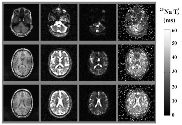
FIG. 1.
T1-weighted axial brain images (8 mm thick) from a healthy volunteer at 7 T (left column), single-quantum sodium images of the corresponding slices at TE = 12 ms and TE = 37 ms (center columns), and parametric map of the selected brain slices (right column). The short TE images were acquired once, while the long TE images were averaged three times, yielding total acquisition time of 63 min (see Materials and Methods section).

