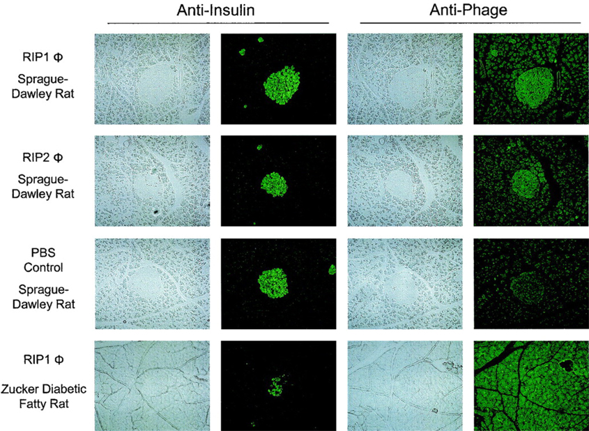
Figure 7.
Distribution of RIP1 and RIP2 phage in the pancreata from Sprague-Dawley or ZDF rats. The phage was injected intravenously into the jugular vein and allowed to circulate for 2 h. Aligned sections are shown staining with anti-insulin or anti-phage antibodies. All panels are at 100 × magnification. Significant accumulation of RIP1 and RIP2 phage in the islets of the Sprague-Dawley rats is visible. No significant accumulation of RIP1 phage is seen within the islet of a ZDF rat. (Copyright © 2005 American Diabetes Association. From Diabetes®, Vol. 54, 2005; 2103–2108. Reprinted with permission from the American Diabetes Association.)

