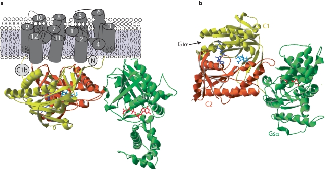
Fig. 1.
Structure of adenylyl cyclase. a Crystal structure of cytoplasmic domains of AC in complex with GTPγ S-Gα, forskolin (FSK) and P-site inhibitor, 2′ 5′ -dideoxy-3′ ATP [100] . Shown are C1 (yellow), C2 (rust), Gs α (green), FSK (cyan), and P-site inhibitor (dark blue). Membrane spans are modeled from the 12-membrane spanning transporters [199] . b Alternate view from cytoplasmic side, showing forskolin and catalytic site more clearly. Interaction site of Giα with C1 domain is indicated by an arrow.

