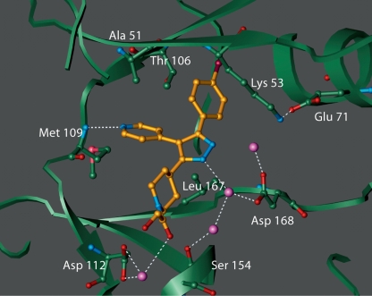
Fig. 2.
Crystal structure of p38α/inhibitor complex. Shown is SC79659 (gold, with nitrogen indicated in blue) which differs from SD0006 only by substitution of the pyridine ring with a pyrimidine and gives the same binding conformation. Binding is at the ATP binding site of p38αlocated at the hinge region between the two lobes of the enzyme. Some of the protein residues that are in close contact with the inhibitor are shown. Also shown are the ordered water molecules (purple spheres). Potential hydrogen bonds are shown in dotted lines. Crystals of p38αcomplex were obtained, and diffraction data were measured, as described in Materials and Methods. The flexible glycine loop has been omitted for clarity.

