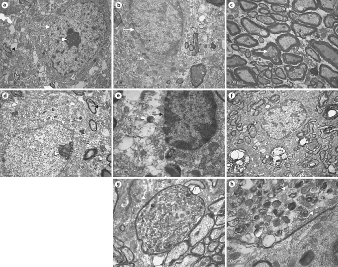
Fig. 7.
Ultrastructural changes in the SNc of lactacystin-treated rats. a The neurons of the right SNc of the rats microinfused with lactacystin in the left SNc showed intact caryotheca (white arrow), limpid nucleoli (arrowhead) and a normal mitochondrial structure (black arrow). b Glial-cell structural morphology was normal and the fine structure of the myelin sheath was limpid in the right SNc of the rats microinfused with lactacystin into the left SNc (c). d Sites ipsilateral to the SNc microinfused with lactacystin: most surviving neurons display apoptotic signs, e.g. caryotheca (white arrow) exhibiting shrinkage, rugosity and partial fragmentation, with intranuclear chromatin condensed by margination (arrowhead), typical of apoptotic cells. In addition, mitochondria (black arrow) in the intracytoplasm were swollen and denatured, even with vacuolization. Several glial cells also showed apoptotic signs (e), and the fine structure of most myelin sheaths became loose and exhibited clear fragmentation (f). Medullary shells also show structural degeneration, e.g. ecphyma swelling and coarsening and a large number of whorls of pronounced electron density (g). h Electron-dense whorl with a core along with whorls with multilamellar fibrils peripherally, which may be protein aggregations (α-synuclein) in the ecphyma.

