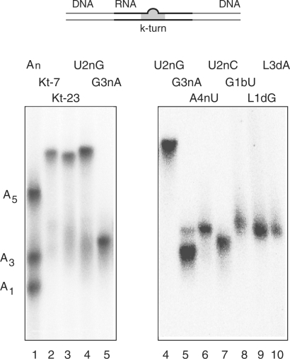
Figure 3.
Electrophoretic analysis of Mg2+-induced kinking of Kt-23 and variants. The scheme (top) shows the 65 bp DNA/RNA/DNA duplex species with a central k-turn sequence used in this analysis. Radioactively [5′-32P]-labelled k-turn species (with or without modifications in the k-turn) were electrophoresed in 13% polyacrylamide gels in 90 mM Tris–borate (pH 8.3) containing 2 mM Mg2+. An-bulge-containing duplexes were made using the same lower strand, hybridized to complement the non-Watson–Crick pairings, and with the three-nucleotide bulge replaced by an A1, A3 or A5 bulge. A mixture of the three species was electrophoresed in track 1, alongside Kt-7 in track 2. The remaining tracks contain: track 3, unmodified Kt-23; track 4, Kt-23 U2nG; track 5, Kt-23 G3nA; track 6, Kt-23 A4nU; track 7, Kt-23 U2nC; track 8, Kt-23 G1bU; track 9, Kt-23 GL12′H; track 10, Kt-23 AL32′H. The samples were electrophoresed in two equivalent gels, with the samples Kt-23 U2nG and G3nA common to both.

