
Figure 2.
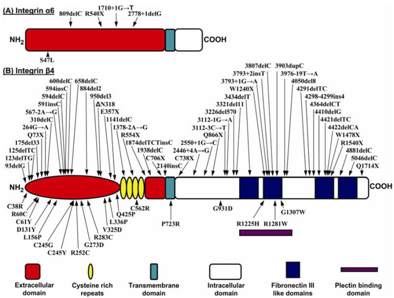
Schematic representation of the domain organizations of the α6 integrin (A) and β4 integrin (B) subunit polypeptides. The color coded explanation of the domains is at the bottom of the figure. The arrows point to the positions of the mutations along the protein structure. Mutations above each of the schematic structure represent premature termination codon-causing mutations, while those below the schematic structures are missense mutations. (Note that the numbering of the mutations may differ from those in the original publications due to the fact that the numbering has been adjusted to conform with the following NCBI database entries: ITGA6, NM-000210; ITGB4, NM-001005731).