FIGURE 1.
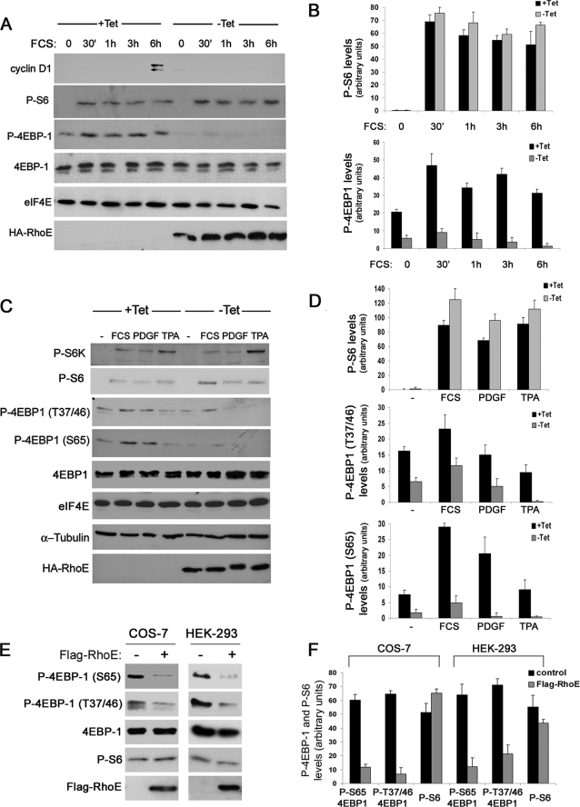
RhoE inhibits growth factor-induced 4E-BP1 phosphorylation. A, RhoE-3T3 cells were starved for 24 h in 0.5% FCS-containing medium in the presence (+Tet) or absence (−Tet) of tetracycline and were stimulated with 10% FCS in the presence or absence of tetracycline and harvested at the indicated time points. The expression levels of the indicated proteins were analyzed by Western blotting with specific antibodies. B, representation of the mean ± S.D. levels of quantified P-S6 and p-4EBP1 in control (+Tet) and RhoE-expressing (−Tet) cells. C, RhoE-3T3 cells were starved as in A, stimulated with 10% FCS, PDGF (1 nm), or TPA (100 nm) for 30 min and harvested. The expression levels of the indicated proteins were analyzed by Western blotting with specific antibodies. D, representation of the mean ± S.D. levels of quantified P-S6 and p-4EBP1 (Ser-65 and Thr-37/46) in control (+Tet) and RhoE-expressing (−Tet) cells. E, COS-7 and HEK-293 cells were transfected with Flag-RhoE and harvested 48 h after transfection. The expression levels of the indicated proteins were analyzed by Western blotting with specific antibodies. F, representation of the mean ± S.D. levels of quantified P-S6 and p-4EBP1 (Ser-65 and Thr-37/46) in mock-transfected (control) and RhoE-transfected (Flag-RhoE) cells.

