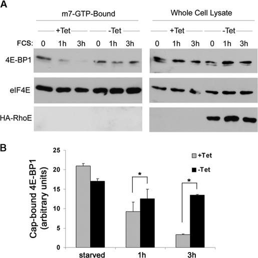
FIGURE 4.
RhoE inhibits 4E-BP1 release from eIF4E in response to mitogenic stimulation. A, RhoE-3T3 cells were starved for 24 h in 0.5% FCS-containing medium in the presence (+Tet) or absence (−Tet) of tetracycline and stimulated for the indicated time with 10% FCS in the presence (+Tet) or absence (−Tet) of tetracycline. Harvested cell lysates were pulled down with m7·GTP-Sepharose as indicated under “Experimental Procedures.” m7·GTP-Sepharose-bound proteins (left panel) and proteins in the input lysate (right panel) were analyzed by Western blotting with the indicated specific antibodies. B, graph represents the mean ± S.D. of quantified 4E-BP1/eIF4E ratio (cap-bound 4E-BP1) from three independent experiments. The differences in cap-bound 4E-BP1 levels between serum-stimulated control (+Tet) and RhoE-expressing cells (−Tet) are statistically significant (Student's t test: *, p < 0.05).

