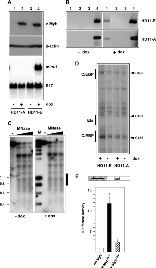
FIGURE 6.
Oncogenic amino acid substitutions abolish the ability of v-Myb to induce chromatin remodeling at the mim-1 enhancer. A, top panels show Western blots of HD11-A and HD11-E cells stained with antibodies against v-Myb and β-actin. The cells were grown in the presence or absence of doxycyclin. The bottom panel show a Northern blot of the same cells hybridized with probes specific for mim-1 and ribosomal protein S17 mRNAs. B, chromatin immunoprecipitation of HD11-E and HD11-A cells grown in the presence or absence of doxycyclin. Immunoprecipitation was carried out using Myb-specific antiserum (lane 1), non-immune serum (lane 2), or no antiserum (lane 3). Lane 4 show input controls. PCR reactions were carried out with primers specific for the mim-1 enhancer. C, micrococcal nuclease digestion of nuclei isolated from HD11-A cells grown in the absence or presence of doxycyclin. The experiment was carried out as described in the legend to Fig. 2B. The black bar on the right marks the position of the mim-1 enhancer. D, DMS footprinting analysis of HD11-E and HD11-A cells. The same region of the mim-1 enhancer was analyzed as in Fig. 3. E, promoter activity of the mim-1 enhancer region in the absence of Myb (white bar) or after co-transfection of expression vector for v-MybREV (black bar) or v-MybAMV (hatched bar). The reporter construct used is shown schematically at the top, and the experiment was carried out as described in Fig. 4C.

