FIGURE 3.
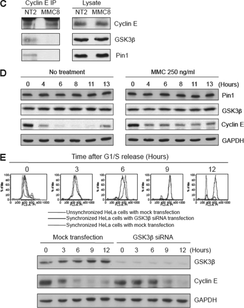
Gradient sedimentation profiles of cyclin E and its binding partners in response to MMC. A, HeLa cells were synchronized and released into regular medium with or without MMC. Lysates were subsequently examined by sucrose gradient sedimentation (left panels). Quantitation of bands is shown in the right panels. L, loading of unfractionated samples. For reference, cell cycle distributions of these cells by FACS analysis are shown in the top panel. B, same as in A except cells were released into MMC. C, immunoblot analysis showing that MMC reduces the interaction between cyclin E and both GSK3β and Pin1 as determined in co-IP experiments. NT2, cells without drug released for 2 h; MMC8, cells treated with MMC (250 ng/ml) and released for 8 h. D, immunoblot showing that the levels of cyclin E, but not Pin1 or GSK3β, are altered in the presence of MMC. E, depletion of GSK3β results in stabilization of cyclin E during S phase. Upper panel shows FACS analysis of HeLa cell treated as indicated. Lower panel shows an immunoblot analysis.


