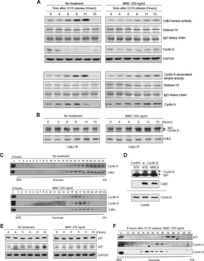
FIGURE 4.
Cdk2 kinase activity is reduced in the presence of MMC. A, upper panels, analyses showing the results of IP-kinase assays of Cdk2 using histone H1 as the substrate. HeLa cells were synchronized and released into regular medium with or without MMC for the indicated times. The top rows show autoradiographs of phosphorylated histone H1. The next two rows shown Coomassie Blue staining of histone H1 and the IgG used for the IP. The bottom two rows show immunoblots of cyclin E and the loading control GAPDH. Lower panels, experiment as described above except that the IP was performed with an antibody to cyclin A. B, immunoblot analysis showing the co-IP of Cdk2 and cyclin E with or without MMC treatment. C, sucrose gradient sedimentation profiles of cyclin A and Cdk2 8 h after release from synchronization with or without MMC. D, immunoblot analysis showing co-IP between cyclin A and Cdk2 8 h after release from synchronization. NT8, nontreated cells at 8 h after release; MMC8, cells released into MMC for 8 h. Control indicates an IP with a nonspecific IgG. E, immunoblot analysis showing levels of p21 and p27 after release into regular medium with or without MMC. F, sucrose gradient sedimentation profiles of p21, cyclin E, and cyclin A at 8 h after release into MMC.

