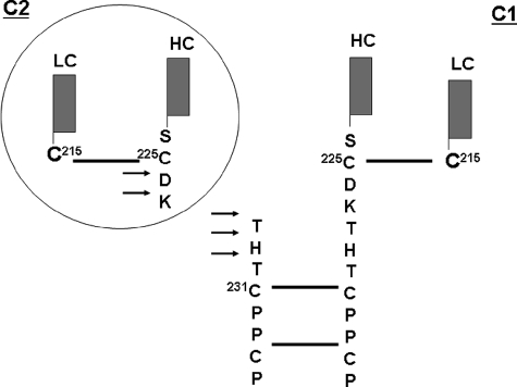
FIGURE 5.
Schematic illustration of the major cleavage sites in the IgG1. H2O2-mediated cleavage of the hinge region, as indicated by the arrows, resulted in the generation of complementary ladders of C- and N-terminal hinge residues (Asp226, Lys227, Thr228, His229, and Thr230) and the release of the C1 and C2 fragments via a one radical per molecule cleavage mechanism (see text for details). Solid lines indicate disulfide bonds.

