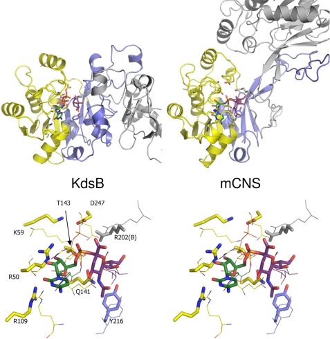
FIGURE 9.
Comparison of E. coli KdsB and the related murine CNS structure. A, KdsB and the related murine CNS structure (PDB code 1QWJ, see Ref. 16) are shown side by side in similar orientations. One monomer is colored coded according to Fig. 4, and the second monomer is shown in gray. Bound ligands and key active side residues are shown in atom-colored sticks (color coding as for Figs. 5–7; murine CNS (mCNS) contains the bound product CMP-5-N-acetylneuraminic acid). B, overlay of the KdsB and murine CNS active site structures. Murine CNS key active site residues and bound ligand are shown in atom-colored sticks, although the corresponding KdsB residues are shown as thin lines for comparison. Active site residues for murine CNS are labeled.

