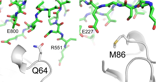
FIGURE 6.
The environment of CRIg residues Gln-64 and Met-86 in the crystal structure of the complex with C3c. CRIg is shown as a white ribbon presentation with the side chains of Met-86 (right) and Gln-64 (left) shown as sticks. C3c is shown in green as stick representations (Protein Data Bank entry 2ICE).

