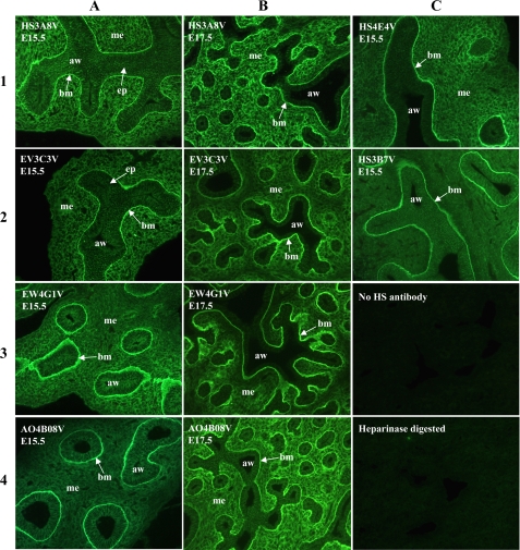
FIGURE 1.
HS phage display antibodies identify distinct HS structures in embryonic rat lung. In E15.5 lungs, HS3B7V exclusively labels epithelial basement membranes and HS4E4V labels basement membranes together with areas of mesenchyme surrounding smaller distal airways. HS3A8V and EV3C3V both label epithelial cells, underlying basement membranes and surrounding mesenchyme in E15.5 lungs; however, at E17.5, staining differs. HS3A8V staining remains strong throughout E17.5 lung mesenchyme, whereas EV3C3V staining becomes more restricted to areas of mesenchyme adjacent to smaller airways. EW4G1V and AO4B08V also show equivalent patterns of staining in E15.5 lungs, strongly labeling epithelial basement membranes and labeling the surrounding mesenchyme more weakly. However, in E17.5 lungs, EW4G1V mesenchymal staining remains weak, whereas AO4B08V mesenchymal staining increases. Immunohistochemistry with individual antibodies is therefore unique, indicating distinct epitopes. E15.5 and E17.5 rat lungs were probed with HS antibodies followed by rabbit VSV-G tag antibody and fluorescein isothiocyanate-conjugated goat anti-rabbit IgG. The negative controls were omission of HS antibody or digestion of HS with heparinase prior to antibody incubation. All of the images are at the same magnification. aw, airway; mes, mesenchyme; epi, epithelium; bm, basement membrane.

