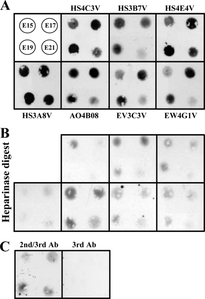
FIGURE 2.
Dot blot analysis of HS epitopes in E15. 5–21.5 fetal rat lung extracts identified differences between in situ and in vitro detection of epitopes. Lung extracts (10 μl) were blotted onto nitrocellulose membrane and probed with HS antibodies as described under “Experimental Procedures.” Bound antibodies were detected via their polyhistidine (His) tag with mouse anti-His-tag IgG, followed by HRP-conjugated sheep anti-mouse IgG. The data are representative of two independent blots. A, the top left panel shows the arrangement of four extracts from E15.5, E17.5, E19.5, and E21.5 fetal lungs. The remaining panels are labeled with the relevant probe HS antibody. B, control data showing background binding after digestion of HS via treatment of lung extracts with a mixture of heparinases I, II, and III (all 5 milliunits/200 of μl extract) prior to blotting onto membrane and probing with antibodies (panels correspond to those in shown in A). C, control probing of blots with secondary anti-His tag and tertiary HRP anti-mouse only (2nd/3rd Ab) or tertiary HRP anti-mouse (3rd Ab) only. Low background immunoreactivity was noted for heparinase-treated lung extract blots; this is likely to reflect reduced enzyme activity in the presence of SDS (data not shown). Ab, antibody.

