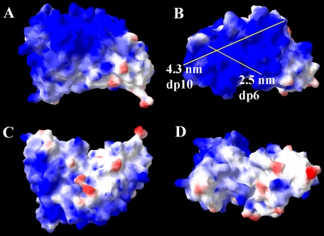
FIGURE 3.
Proposed model and surface electrostatic potential of HS4E4V from four perspectives. A large proportion of basic residues are observed on the surface of the antibody, which will interact with HS and heparin. Two possible HS-binding axes and their lengths are shown. The structure of HS4E4V is extremely similar to HS3A8V, EV3C3V, EW4G1V, and AO4B08V, shown in supplemental Figs. S1–S4. Regions of positive surface charge are represented in blue, and regions of negative charge are in red, whereas neutral areas are white.

