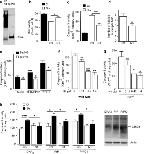
FIGURE 1.
Recombinant N1 fragment protects cells against staurosporine-induced apoptotic cell death in HEK293 cells and mouse primary cultured cortical neurons. a, recombinant N1 fragment was produced in E. coli and purified as described under “Materials and Methods.” Secreted N1 was immunocaptured from HEK293 supernatant as described under “Materials and Methods,” analyzed by 16.5% Tris/Tricine SDS-PAGE, and revealed by immunoblotting with SAF32. b–d, 1 μm recombinant N1 or an equivalent volume of supernatant produced after thrombin digestion of GST-Sepharose beads (KG) was applied to HEK293 cells. After 4 h of incubation, cells were treated again with N1 or KG, before being challenged with staurosporine (2 μm, 16 h) and processed for XTT measurement (b), caspase-3 activity determination (c), or TUNEL labeling (d). Bars in b and c, means ± S.E. of 4–6 independent determinations with two technical replicas. Bars in d, means ± S.E. of the number of labeled nuclei in 10 independent optical fields. e, caspase-3 activity in mock-transfected (Mock) HEK293, 3F4MoPrP, or C1-expressing cells treated with N1 or KG as detailed above. f and g, primary cultured cortical neurons were obtained from the indicated embryonic day 14 mouse embryos as described under “Materials and Methods” and treated as detailed above with increasing concentrations of N1, prior to staurosporine challenge (2 μm, 16 h) and measurement of caspase-3 activity. Bars, means ± S.E. of 3–4 independent determinations carried out in duplicate. h, caspase-3 activity measured in primary cultured mouse cortical neurons transfected with the cDNA of full-length PrP (PrP), C1 (PrPC1), or parental plasmid (DNA3). Forty-eight h post-transfection, cells were incubated with N1 (1 μm, 4 h) or an equivalent volume of KG. Cells were treated again with N1, challenged with staurosporine (2 μm, 16 h), and processed for caspase-3 activity determination. Bars, means of three independent determinations carried out in duplicate. Right, a representative Western blot using SAF61 antibody of PrPc and C1 expression in cortical neurons after nucleofection. *, p < 0.05; **, p < 0.001; ***, p < 0.0001; ns, not significant; IP, immunoprecipitation.

