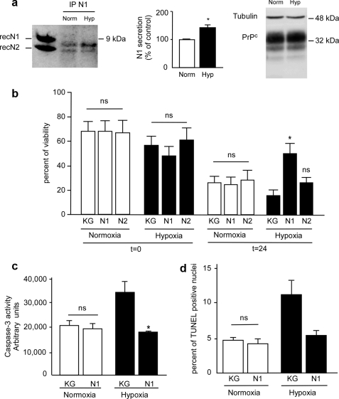
FIGURE 6.
N1 protects rat RGC against OGD. a, RGC were subjected or not to OGD for 2 h (see “Materials and Methods”) and then returned to complete medium for 24 h. Medium was collected, and N1 was immunoprecipitated (IP) with the monoclonal antibody SAF32 and analyzed by 16.5% Tris/Tricine SDS-PAGE and Western blotting using SAF32. RecN1 and RecN2 correspond to the migration pattern of recombinant N1 and N2 (150 ng) analyzed under the same conditions. Bars correspond to the densitometric analysis of N1 and are expressed as a percentage of control N1 recovered under normoxic conditions. Values are the means ± S.E. of three independent experiments. b, 7 days after dissociation, RGC were treated with N1 (2 μm, 4 h) or an equivalent volume of KG, cells were then retreated and placed under normoxic or hypoxic conditions as in a, viability was determined just before return to complete medium or after 24 h by measurement of release of lactate dehydrogenase in the culture medium as described under “Materials and Methods.” c, caspase-3 activity was measured 24 h posthypoxia. d, cells plated on coverslips were treated as in a and processed for TUNEL staining. Bars represent the means ± S.E. of three independent determinations carried out in duplicate. *, p < 0.05.

