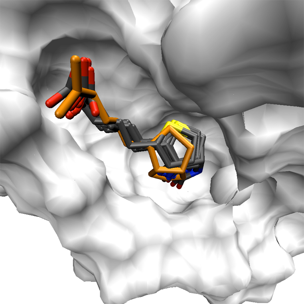
Figure 10. The minor conformation of the biotin tail fills a similar volume as the major conformation.
While the alternate, minor conformation of the biotin tail involves simultaneous changes in two dihedral angles, it occupies much the same volume as the conformation observed in X-ray structures. Furthermore, the carboxylate group on the biotin tail sits in a similar orientation for both conformations of the tail, maintaining hydrogen bonds to Ser88 Oγ and Asn49 N. Eight conformations of the biotin are shown in licorice, superimposed against the white surface representing a streptavidin monomer. One of the biotins, highlighted in orange, exhibits the minor tail conformation. The streptavidin core backbone atoms were aligned to superimpose the various biotin conformations.

