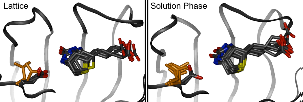
Figure 15. Motion of Asp128 as it breaks its hydrogen bond to biotin in the cryo-solvated lattice and solution phase simulations.
Eight conformations of the biotin binding site are superimposed on one another (positions of β-barrel core backbone atoms were used for alignment) in each frame. A backbone trace of the streptavidin protein is shown as a black ribbon, while the biotin ligand and Asp128 side chain are shown in licorice. If the the hydrogen bond between Asp128 Oδ and biotin N1 is broken, the Asp128 side chain is highlighted in orange. The motion of Asp128 during hydrogen bond cleavage is similar in both simulations.

