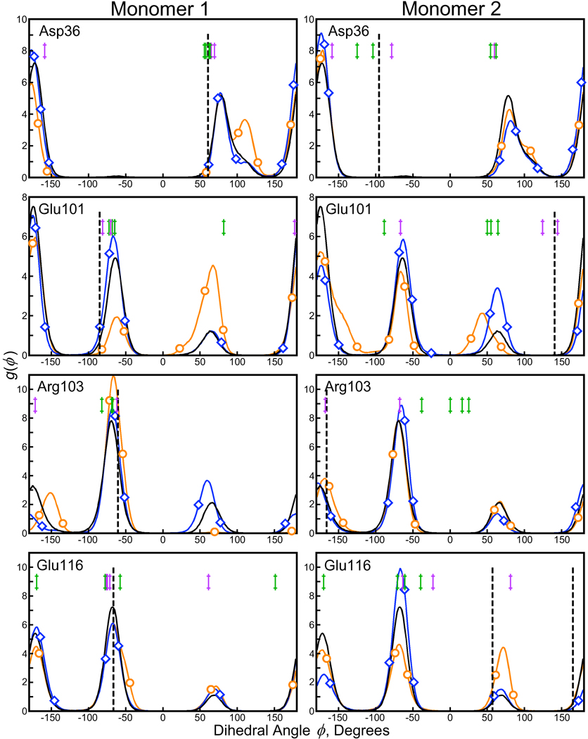
Figure 7. χ1 distributions for selected residues making lattice contacts in the 1MK5 structure.
Experimental value(s) for each dihedral angle as well as distributions obtained from solution phase and crystal lattice simulations are plotted according to the color scheme of Figure 6. Residues presented in this plot diverged from the results in the 1MK5 structure, but isomorphous crystals grown and analyzed under similar conditions (1DF8, 1LUQ, and 2F01) showed some diversity in the same dihedral angles (denoted by purple marks in each panel). When X-ray data was collected at room temperature, the diversity increased even further (structures 1LCV, 1PTS, 1SLG, and 1SRI; green marks in each panel). The differences between simulation and experiment are not necessarily errors; additional simulations with longer trajectories may help provide more certainty in the populations of each χ1 angle.

