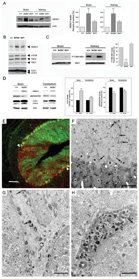
Figure 7. Herc1 expression and functional analysis.
Brain (A–D), kidney (A,C) and cerebellum (D) homogenates from wild-type (+/+), heterozygous (+/tbl) and homozygous (tbl/tbl) mice were analyzed by Western-blot using specific antibodies against the indicated proteins. HERC1 (in brain and in kidney), P-T389-S6K1 (in kidney), p62/SQSTM1 and LC3-II (in brain and in cerebellum) levels were quantified (n = 4–9) and expressed as the mean±s.d. of percentage of respective control. ** p<0.01,*** p<0.001. (E) Parasagittal section of a 2-month-old tbl/tbl cerebellum double immunostained with anti-calbindin D28k antibodies to visualize PC (green) and anti-LAMP-1 (red) to identify lysosomes and autophagosomes. The arrowheads point to an area of the cerebellar cortex almost devoid of PC. In this area, LAMP-1 positive puncta are numerous, testifying for the degeneration of PC. (F) Parasagittal 1µm-thick plastic section stained with toluidine blue, illustrating four PC somata (asterisks). The arrows point to dense cytoplasmic inclusions accumulated in the cell bodies and proximal dendrites, particularly at their branching points. The arrowhead points to the first Ranvier node of a PC axon initial segment, which looks normal. (G,H) Electron-micrographs made from the same mouse showing two profiles of the proximal dendritic compartment. The one in (G) illustrates a branching zone in an early stage of the autophagic process. The dendrite contains some vacuoles bound by a smooth double membrane (arrows), enclosing whorls of membrane-like elements, and dense debris (asterisks) suspended in an electronlucent matrix. The other dendritic profile (H) corresponds to a more advanced stage in autophagy, characterized by the occurrence of extremely numerous single membrane bound vacuoles corresponding to autolysosomes, some of them of large size (asterisk). The arrow points to a giant spine emerging from the dendrite and postsynaptic to several parallel fibers varicosities. Scale bars: the bar is equal to 100 µm in (E), 24 µm in (F), 1.2 µm in (G), and 1.5 µm in (H).

