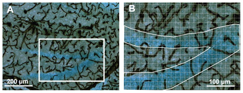
FIGURE 2.
Blood vessels in the hippocampus. (A) A representative section stained with an antibody against collagen IV with diaminobenzidine as the chromogen, combined with a light Nissl stain to visualize blood vessels in and around the dentate gyrus (×100 total magnification). (B) The same section at ×200 total magnification with grid overlaid and region outlined to illustrate the method for estimating area fraction covered by vessels in the granular layer. Twelve or more such pictures per animal were analyzed to count the number of vertices intersecting with vascular tissue within defined brain regions. [Color figure can be viewed in the online issue, which is available at www.interscience.wiley.com.]

