
Fig 1.
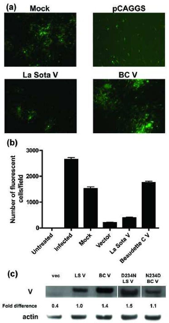
BC V exhibits a four-fold greater ability than La Sota V to rescue growth of NDV-GFP. (A) DF1 cells were mock-transfected, or transfected with empty pCAGGS vector, La Sota V or BC V. After 24 h, the cells were infected with NDV-GFP virus and examined for fluorescence 24 h later. (B) The growth of NDV-GFP virus was quantitated by counting the number of fluorescent cells from three different fields. The average values and standard deviations (error bars) are shown for one experiment out of a total of four. The absolute numbers of fluorescent cells vary from one experiment to another but the relative activities are consistent. (C) DF1 cells were transfected with wt or mutant V plasmids. After 24 h, lysates were prepared and Western blot was performed using V18 antiserum. V protein levels standardized to actin were determined by densitometry and are expressed relative to wt (set at 100%). These data represent one out of at least two experiments.