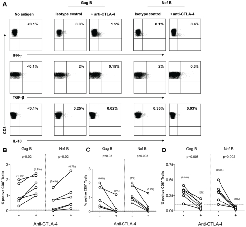
Figure 3. CTLA-4 blockade decreases TGF-β and IL-10 expression by HIV-specific CD8+ T Cells.
PBMC (n = 6) were stimulated with HIV peptides in the presence of anti-CTLA4 (or isotype control), then stained with anti-IFN-γ FITC, anti-TGF-β PE, anti-IL-10 APC, anti-CD3 AmCyan, anti-CD4 PerCP CY5.5, anti-CD8 PE CY7, and analyzed by flow cytomerty. Samples were first gated on the CD3+/CD8+ lymphocyte population then the percent of TGF-β, IL-10, and IFN-γ positive cells were determined. Results were expressed as percent of HIV-specific CD8+ T cells expressing TGF-β, IL-10, or IFN-γ after subtraction of the back ground. (A) Representative plots of HIV-specific CD8+ T cells expressing TGF-β, IL-10, or IFN-γ in the presence or absence of anti-CTLA-4. (B-D) Dashed line represents the cutoff for significant TGF-β (B), IL-10 (C), and IFN-γ (D) expression. Percentages in between brackets are median values. The two dots joined by a line represent the values obtained from the same individual and analysis was performed by paired t-test.

