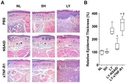
Figure 2. Histological responses to pharmacotherapy.
Tail sections were harvested 16 mm from the base of the tail, stained with hematoxylin/eosin, and examined by light microscopy. (A) Representative histology. Specimens from normal control (NL) mice, sham surgery control (SH) mice, and lymphedema (LY) mice treated with either PBS, ketoprofen (NSAID), or the TNF-α inhibitor sTNFR-1. Untreated LY show hyperkeratosis, epidermal spongiosis and edema, irregularity of the epidermal/dermal junction, elongation of the dermal papillae, and a 2- to 3-fold expansion of tissue between the bone and the epidermis. There are numerous dilated microlymphatics and increased cellularity, including a large infiltration of neutrophils. Treatment with NSAID normalizes these pathological findings whereas treatment with sTNFR-1 exacerbates the pathology. (B) Quantification of epidermal thickness (ET). Changes in ET are expressed as a percentage of the average ET of NL. ET of NSAID-treated lymphedema (LY-NSAID) mice was significantly reduced compared to untreated LY mice (P<0.0005) and were not significantly different than NL or SH control mice. ET of sTNF-R1 treated lymphedema (LY-sTNF-R1) mice was significantly increased compared to untreated LY mice (P<0.05). *P<0.05 vs LY, † P<0.0005 vs LY-NSAID.

