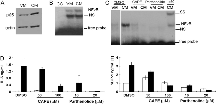
Fig. 1.
NF-κB activation in osteoblasts exposed to breast cancer cell-conditioned medium (BCCM). MC3T3-E1 were cultured and differentiated for 2 weeks before stimulation with 50% BCCM for different times. (A) p65 translocation was detected by western blot using nuclear extracts prepared from MC3T3-E1 1 h after treatment with BCCM. (B) NF-κB gel mobility shift analysis (EMSA) of samples prepared as in (A); CC, cold oligonucleotide competitor; NS, non-specific binding; SS, super-shift with anti-p50. (C) Inhibition of NF-κB binding. Osteoblasts were pretreated with inhibitors CAPE for 2 h or with parthenolide for 1 h before addition of BCCM for 1 h. NF-κB–DNA-binding activity was detected by an EMSA p50 supershift. (D) IL-6 and (E) MCP-1 production by osteoblasts treated with NF-κB inhibitors CAPE for 2 h or with parthenolide for 1 h before incubation with BCCM for 4 h. DMSO (0.2%) treatment was used as control. Cytokines in the culture media were detected by enzyme-linked immunosorbent assay. Open bars indicate treatment with VM; closed bars indicate treatment with BCCM. All experiments were performed twice and each sample was assayed in duplicate.

