
Fig. 6.
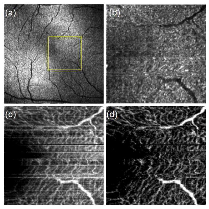
OCT summation images over the foveal region of the retina. (a) 3mm x 3mm intensity summation image identifying approximate location of 1mm x 1mm scan area. (b) 1 mm x 1 mm intensity summation image. Motion contrast retinal summation images (c) before and (d) after additional noise removal..