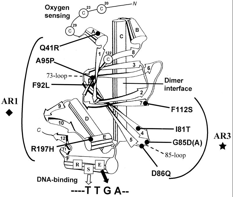
Figure 2.
Predicted structure of an FNR monomer based on that of CRP (14) showing the positions of amino acid substitutions that confer a hemolytic phenotype on E. coli K12. Also shown are: the helix–turn–helix motif (αE-αF) in the DNA-binding domain; key amino acid to base pair (protein–DNA) interactions; the essential cysteine ligands of the [4Fe-4S] cluster; and previously identified activating regions, the 73-loop in AR1 (13) and the 85-loop in AR3 (13).

