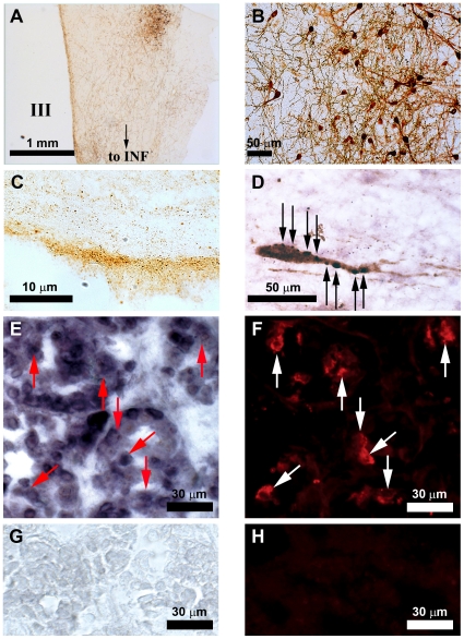
Figure 1. GnIH immunoreactive neurons in the human hypothalamus and GnIH receptor mRNA in the human pituitary.
(A) Coronal section of adult human hypothalamus showing GnIH immunoreactive (-ir) neurons clustered in the dorsomedial region of the hypothalamus with their fibers extending to the infundibulum (INF). III, third ventricle. Bar, 1 mm. (B) Higher magnification of GnIH-ir neuronal cell bodies. Bar, 50 µm. (C) Sagittal section of the median eminence showing dense population of GnIH-ir fibers within the external layer. Bar, 10 µm. (D) GnIH-ir axon terminal like structures (stained in purple, indicated by black arrows) in close proximity to a GnRH-ir neuronal cell body (stained in brown) in the preoptic area. Bar, 50 µm. (E and F) In situ hybridization of human GnIH receptor (GPR147) mRNA in the human anterior pituitary (E) in combination with immunocytochemistry for luteinizing hormone (LH) (F). Red arrows in (E) and white arrows in (F) in equivalent positions show cells which express both GPR147 mRNA and LH. Bars, 30 µm. (G and H) In situ hybridization using sense RNA probe (G) and immunocytochemistry without LH primary antiserum (H) served as controls. Bars, 30 µm.

