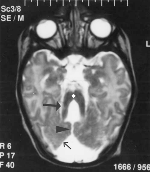
Figure 3).
Axial T2 weighted image shows dysgenetic cerebellar hemispheres (small arrow) with a prominent vermian cleft (arrowhead) and narrowed isthmus of the mid brain (diamond). The superior cerebellar peduncles bilaterally appear enlarged and horizontal, resulting in characteristic ‘molar tooth appearance’ (big arrow)

