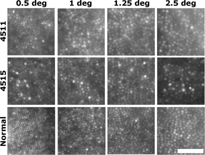
Fig. 1.
Visualizing cone mosaic disruption in the C203R retina. Shown are images from the right eyes of subject 4511, 4515, and a normal control from 0.5°, 1°, 1.25°, and 2.5° temporal to the fovea. Retinal magnification estimates are 288 μm/degree (4511), 281 μm/degree (4515), and 298 μm/degree (normal control). [Scale bar, 50 μm (≈10.5 arcmin).]

