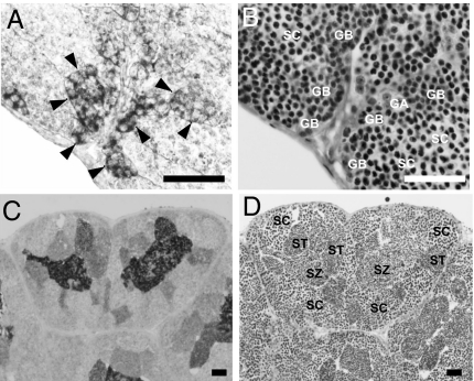
Fig. 2.
Cellular localization of eel trypsinogen in the testis. (A and B) Eel testis at 12 days post-hCG injection. (C and D) Eel testis at 18 days post-hCG injection. (A and C) were assessed by histochemistry using a specific anti-eel trypsinogen antibody. (B and D) show serial sections of A and C, respectively, stained with hematoxylin and eosin. Type A and early type B spermatogonia (GA), late type B spermatogonia (GB), spermatocytes (SC) spermatids (ST), spermatozoa (SZ), and enlarged Sertoli cells (arrowheads) are also shown. (Scale bar, 50 μm.)

