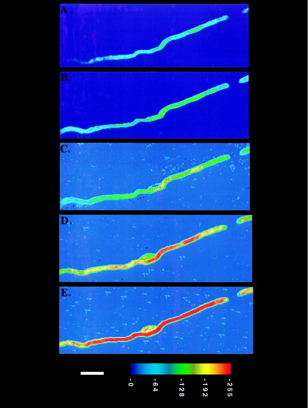
Figure 1.
Time course of myelin formation by a single Schwann cell on an axon. C5-DMB-hexadecanoic acid (0.75 μM) was added directly to the coculture and its incorporation was monitored for 30 hr. The coculture was placed in a temperature-controlled (35°C) perfusion chamber and viewed with a fluorescence digital imaging microscope. Changes in the fluorescence intensity of the internode were observed at 5 hr (A), 10 hr (B), 13 hr (C), 16 hr (D), and 22 hr (E) after labeling. The cell body of the Schwann cell is visible in the middle of the internode in the last three frames. The background fluorescence attributable to the node of Ranvier is detected in the upper right corner along with the tip of the adjacent internode. The axon is not visible. The applied pseudocolor scheme and a 10-μm bar appear at the bottom.

